Technical Specification of the Multimode SPM
This page gives a very basic specification and description of the instrument and contains links to the relevant sections of the manual. It is split into 3 sections:
Hardware
The NanoScope MultiMode AFM performs the full range of SPM techniques to measure surface
characteristics including topography, elasticity, friction, adhesion, magnetic
fields, and electrical fields. This microscope is ideal for smaller samples (maximum
of 15 mm in diameter and 5 mm thick) for larger samples the Park Scientific Instruments Combined STM-SA1 and SFM-BD2 is availiable. The multimode allows both conductive and
non-conductive samples to be studied. It supports contact, non-contact, Tapping
Mode, lateral force, magnetic force, and electric force techniques, as well as
STM and electrochemistry. It is equipped with an Extender Electronics Module
that enhances magnetic and electric field imaging.
The innovative, compact
and rigid construction of the hardware give the MultiMode SPM the mechanical
stability and low noise needed for high resolution (it is capable of true
atomic resolution). It is thus possible
to easily acquire images on both the atomic and macroscopic scales. The
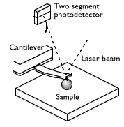
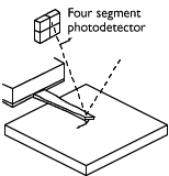
hardware includes an optical detection head, TappingMode (air) Cantilever
Holder, TappingMode Fluid Cell, Low-Current STM Converter, 2 scanners, and
microscope base. An Optical Viewing System consisting of 450x optical viewing
system with optical microscope, colour CCD camera, and colour monitor; provide
vertical optical view of tip and sample. The Extender Electronics Module is
also available: that contains phase and frequency detection hardware for Phase
Imaging, MFM and EFM; required for Surface Potential measurements and
recommended for all applications
The MultiMode SPM and Nanoscope
IIIb control system are designed to provide superior scanning control combined
with ease of use and reliability. The compact, rigid construction and low noise
level make it the ideal microscope for atomic scale imaging. The short
mechanical path length between tip and sample produces the high resonance
frequency required for the fast scan rates that are essential for atomic
resolution. In addition, careful choice of critical components ensures high
thermal stability.
The controller provides 16-bit resolution
on all three axes (x, y, and z scanner drives with ±220V range), with three
independent 16-bit digital-to-analog converters (DACs) in x and y
for control of the scan pattern, scaling, and offset. This configuration
provides 16-bit resolution of the lateral scanning motion at any scan size, and
the ability to perform atomic resolution imaging throughout the full lateral
range of the scanner. The patented digital feedback is governed by integral and
proportional gain controls, providing immediate response to scanning parameter
changes.
The MultiMode
can scan up to 100µm laterally (x and y axes) and 10µm vertically
with the J scanner and up to 10µm laterally (x and y axes) and 3.5µm
vertically (z axis) with the E scanner. The scanner calibration and
linearization are maintained by software control, providing the user with easy,
direct access to the calibration parameters of the scanner. The scanner
maintains its calibration regardless of the scan size, offset, or direction.
The patented design of the 12µm and 130µm lateral range scanners consists of a
“hard” piezoelectric material for the vertical movement, which minimizes the
effects of nonlinearity and hysteresis while maintaining calibration throughout
the full vertical range.
Scanning Techniques
The complete
range of Atomic Force Microscopy (AFM) and Scanning Tunneling Microscopy (STM)
techniques is available with the MultiMode SPM:
Software
The microscope
is controlled by powerful but easy to use control, image analysis and
presentation software, that also contains powerful algorithms for the
measurement and presentation of your results. You can view your images in two-
and three-dimensional representations, with a variety of color schemes with the
ability to design your own. You can also analyze your images with a variety of
algorithms, including:
Several image modification algorithms are also provided, including:
With the “Auto Program” feature, an off-line macro routine can be easily created to perform a series of analysis and modification steps with or without the user present. The results are stored in files which can then be printed or exported as ASCII files for use with other software packages. Similarly, NanoScope image files can also be exported and imported as TIFF or ASCII files for use with other
software.
Consumable Costs
| Contact Mode Probe |
Ł7.50 |
| Tapping Mode Probe |
Ł50 |
For more details on these probes click here.
Back to the instruments page


