| Mirror Speed (in cm/sec) | Associated Laser Modulation Frequency (in KHz) | IR Modulation Frequencies (in Hz) at 400 cm-1 | IR Modulation Frequencies (in Hz) at 4000 cm-1 |
| 0.16 | 5 | 126 | 1260 |
| 0.64 | 20 | 504 | 5040 |
| material | Germanium | KRS-5 | Silicon | Zinc Selenide | Diamond |
| Useful range: | |||||
| maximum/cm-1 | 5500 | 20000 | 8300 | 20000 | 4500 |
| minimum/cm-1 | 830 | 400 | 70 | 650 | 33 |
| Refractive Index at 1000cm-1 | 4 | 2.37 | 3.4 | 2.4 | 2.4 |
| Penetration Depths: | |||||
| incident at 45o/microns | 0.65 | 1.73 | 0.81 | 1.66 | 1.66 |
| incident at 60o/microns | 0.5 | 1.06 | 0.61 | 1.04 | 1.04 |
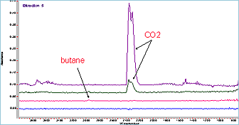
| Strong Absorption/cm-1 | 1500-360 | 2500-1667 | |||
| Incompatible with | Clean with | |
| Germanium | Hot H2SO4 and aqua regia | Alcohol. Acetone, water |
| KRS-5 | complexing agents/water | MEK |
| Silicon | HF + HNO3 | Alcohol. Acetone, water |
| Zinc Selenide | Acids Strong alkalis | Alcohol. Acetone, water |
| Diamond | K2Cr2O7, conc H2SO4 | Alcohol. Acetone, water |